Under the skin: teaching the body

Under the skin: teaching the body


Through history to the present day, artists, anatomists and doctors have both competed and collaborated over the possession, interpretation and depiction of our flesh and bones. Along with photographers, filmmakers, printers, scientists and sculptors they have developed diverse and diverging approaches to representing the layers of our physical selves. Opening a profound conversation on what it means to inhabit, and own, a human body.

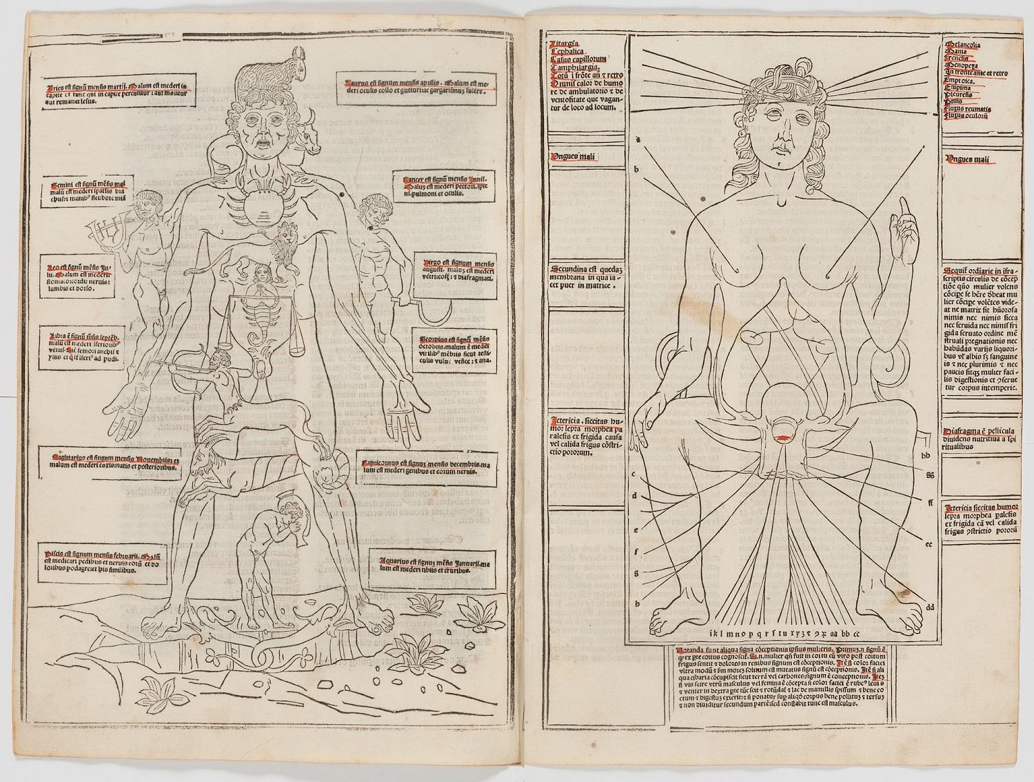
Diagram of a male body and signs of the zodiac on the left, diagram of the female body on the right.

Zodiac man and female anatomy

The male body shown with its corresponding signs of the zodiac, and female anatomy, in Fasciculus medicine: similitudo complexionum & elementorum. Text attributed to Johannes de Ketham, artists unknown, published Venice, 1500.

This short compendium describes the key texts required by medical students at the end of the 15th century. The first edition, which appeared in 1493, was the first printed book to include anatomical illustrations.

The woodcut of the ‘zodiac man’ on the left shows the conceptual relationships between the 12 signs of the zodiac and different parts of the human body, a connection between earth and the heavens. The woodcut on the right, which shows the anatomy of the female body, has a very different artistic style, suggesting the two images were copied from models from different traditions.

View the catalogue record for Fasciculus medicine: similitudo complexionum & elementorum or Explore the complete book online

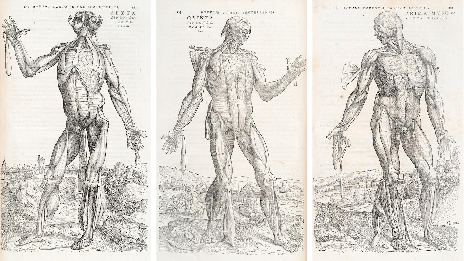
Three image of a male body with partially flayed skin, showing the muscles.

Muscles of the male human body

Muscles of the male human body (front), in De humani corporis fabrica libri septem. Text and dissection by Andreas Vesalius, drawing attributed to Jan Stephan van Calcar, woodcut artist unknown, published Basel, 1543.

Vesalius’ De humani corporis is one of the most famous books in the history of medicine and art. It shows the human body with a previously unknown level of detail, accuracy and artistic flair.

The book uses woodcut illustrations with an extraordinary level of skill. The wood blocks were very subtly carved, with fine lines combined to create three-dimensional form, texture, and movement.

The images – which include dissection views and full figures – were innovative in their detail and poses. The images became frequently-copied standard models in the centuries after their publication.

View the catalogue record for De humani corporis fabrica libri septem or explore the second edition (1555) online

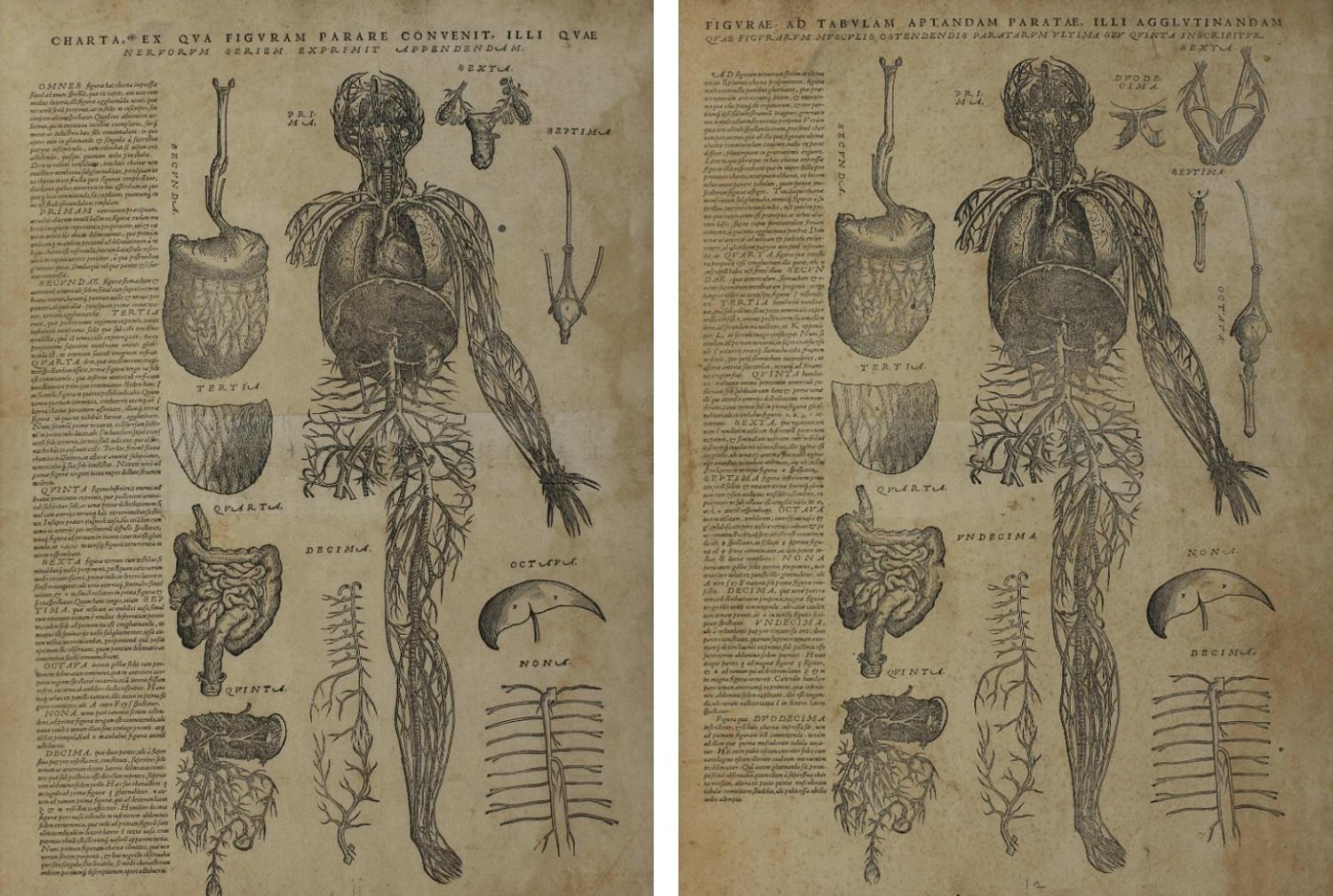
Anatomical drawing of the blood vessesls of the body with organs surrounding the main body. Two versions: one male and one female.

Female and male internal anatomy

Female and male internal anatomy, in De humani corporis fabrica librorum epitome. Andreas Vesalius, published Basel, 1543.

This is a student edition of Vesalius’ masterwork De humani corporis fabrica, published in the same year as the first full edition. It condenses the original 650-page text down to the essentials, and includes the key illustrations.

These images were meant to be cut up and pasted together by the owner as a lift-the-flap diagram.

View the catalogue record for De humani corporis fabrica librorum epitome or explore the complete book online

Download a kit to make your own anatomical flap model

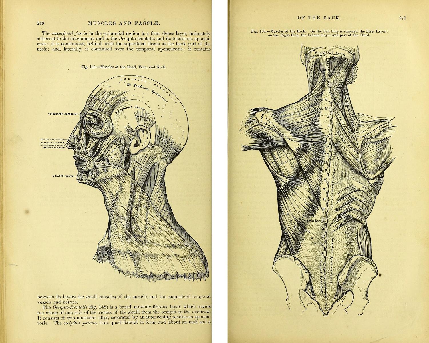
Muscles of the head, face, neck, and back

Muscles of the head, face and neck, and muscles of the back in Anatomy: descriptive and surgical. Dissection by Henry Gray and Henry Vandyke Carter, drawing by Henry Vandyke Carter, published Philadelphia, 1867 (second American edition). 

Gray’s anatomy is probably the most famous medical textbook. It has never been out of print since it was first published in 1858, and today it has reached its 41st edition.

The success of the book can be partly attributed to surgeon and artist Henry Vandyke Carter, who drew the illustrations for the first edition. Vandyke Carter innovatively overlaid descriptive text onto the images themselves, making it easier for the student to connect description and image without having to turn pages.

View the catalogue record for Anatomy: descriptive and surgical. or explore the whole book online.


Eric Beck talks about the lunch time post-mortems that he used to attend

Eric Beck (1934–2019) was a consultant physician and gastroenterologist who trained in medicine London in the 1950s.

'The other thing about the Hammersmith which was a habit which I then saw in other places was after you’d had your lunch you went to the post-mortem room to see, as it were, the corpse of the day. And this was a, a very effective bit of teaching. That the pathologist would, would be doing an autopsy much in advance of the audience coming and the houseman would present the history of the case of the person who died and the pathologist would then demonstrate what he had found which usually fitted in reasonably well with the ante-mortem diagnosis but occasionally revealed some very striking conflicts. And again it made the point, you know, the importance of teamwork and people talking to each other and how much you learned from your colleagues.'

Listen to the full interview for Eric Beck online or Explore more oral history recordings about anatomy and imaging

Barbara Bannister remembers her anatomy teachers at the Royal Free Hospital

Barbara Bannister (b.1948) is a fellow of the RCP who specialised in infectious and tropical diseases. She studied medicine at the Royal Free Hospital in London.

'And there was an even more redoubtable elderly professor who’d retired and she was very strict and she used to tease the boys relentlessly if they couldn’t recite the anatomy of the female pelvis. And there was a lady surgeon who was very good on anatomy of course and very good on how to crash your car without breaking your hips and would sit at the side of the body, because we had a whole body in those days and we had to work on it and look after it because of course it had been donated for us, and she would sit in the chair and raise her arms to both sides shouting "if I am the uterus and these are my tubes."'

Listen to the full interview with Barbara Bannister online or Explore more oral history recordings about anatomy and imaging

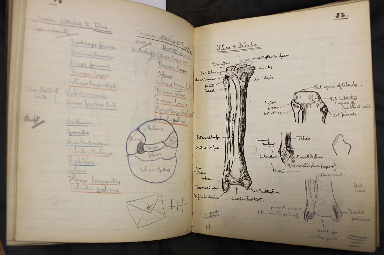
Exercise book with hand drawn illustrations of bones of the lower leg.

Bones of the lower leg

Bones of the lower leg, in Notebook of anatomical notes and drawings. Francis Martin Rouse Walshe, October—December 1904.

Neurologist Francis Walshe made these drawings in pencil, ink and coloured pencil during his medical studies at University College Hospital, London.

Walshe combines realistic three-dimensional drawings with careful shading, with cross-sections showing internal structures.

View the catalogue record for Walshe's notebook

Curious anatomy's : the anatomical tables of the RCP

The RCP museum holds a rare set of six anatomical tables. They are amongst the oldest surviving human anatomy preparations in Europe. The tables are visually spectacular. They show the recognisable form of the human body laid out flat, as if illustrating a book. The nerves, veins and arteries on display were dissected at the University of Padua’s famous anatomy theatre in the 17th century, and skilfully arranged on varnished wooden panels.

One of only two sets known to exist, the tables were probably created as teaching aids for medical students, or as experiments in preserving human tissue. Their production would have required a huge amount of time and extraordinary skill. We believe that the English physician, anatomist and diplomat Sir John Finch (1626–82) brought the tables to England. In 1823 they were discovered in a country house library and presented to the RCP. Unfortunately, much about their origin and history remains a mystery. 

Discover more about the anatomical tables or view the catalogue record for the anatomical tables.

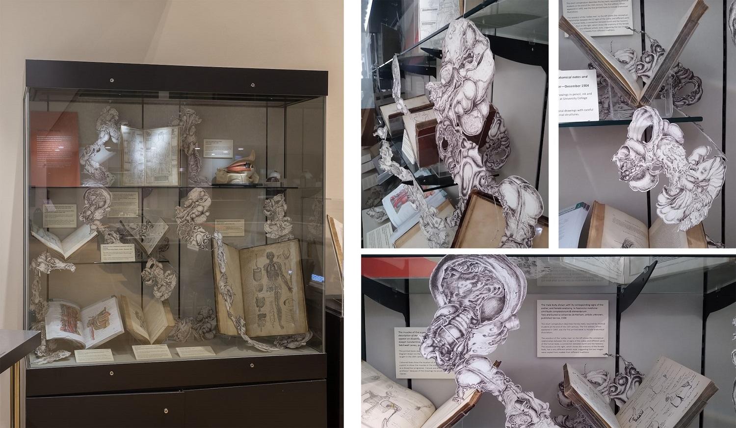
Composite drawings of anatomical illustrations, intertwined with a museum display of open books.

Impossible pathologies

Impossible pathologies. Lucy Lyons, pen on paper, 2017.

These drawings are composites based on original drawings by physician Robert Hooper (1773–1835). They were copied and reprinted, and then assembled into new, composite forms, which are medically impossible and seem to escape the confines of the body.

The focus on hand-drawing reflects the long history of drawing as a learning and teaching tool in the dissecting room.


Part of the exhibition 'Under the skin: anatomy, art and identity'. Explore further: